Understanding PMS and Pregnancy: What You Need to Know
January 23 2025Dermoscopy – Your Window into Skin Health
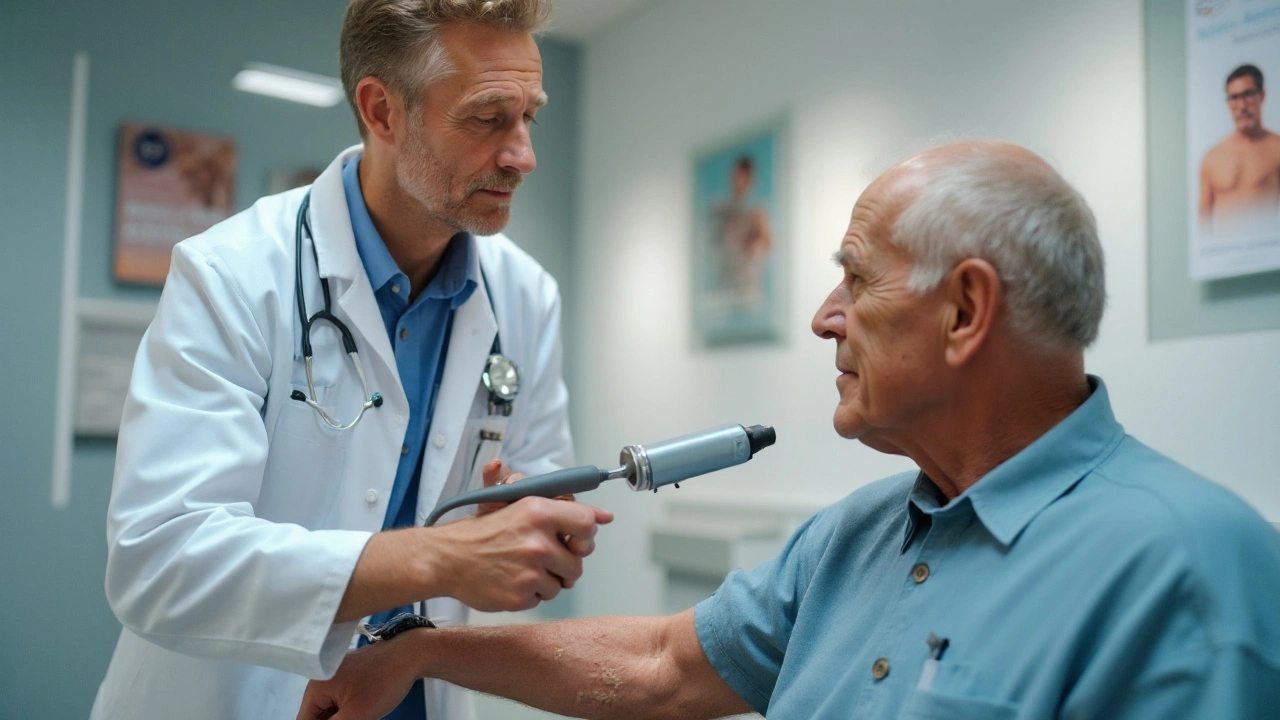
When working with dermoscopy, a non‑invasive imaging technique that magnifies the surface of the skin to reveal hidden structures. Also known as skin surface microscopy, it enables doctors to tell the difference between harmless spots and potentially dangerous growths. Skin lesions are any abnormal changes in color, shape or texture of the skin become much clearer under a dermatoscope, and the method is especially valuable for spotting melanoma, the deadliest form of skin cancer that often looks like a regular mole at first glance. In short, dermoscopy encompasses detailed lesion assessment, requires a handheld dermatoscope, and influences early cancer detection.
Key Tools and Practices That Boost Dermoscopy Effectiveness
The most common handheld dermatoscope, a lightweight, magnifying device with polarized or non‑polarized light gives clinicians a 10x to 30x view of pigment patterns, vascular structures and surface texture. Modern models often connect to smartphones, turning the phone into a digital archive and enabling teledermatology, remote skin consultations where images are shared with specialists for a second opinion. This synergy expands access to expert evaluation, especially in rural areas where a dermatologist might not be nearby.
Another important related entity is digital dermoscopy, software that stores, compares and analyzes images over time. By tracking changes, doctors can apply the “ABCD” rule (Asymmetry, Border, Color, Diameter) more accurately and decide whether a biopsy is needed. The practice of skin cancer screening, routine checks for suspicious lesions in at‑risk populations often relies on these digital tools to catch melanoma at an early, treatable stage.
For patients, understanding dermoscopy basics empowers better self‑care. Knowing that a new, changing, or itchy mole warrants a closer look can prompt an early appointment. Likewise, being aware that dermoscopy can spot subtle variations—like a streak of color or an irregular border—helps set realistic expectations about what a dermatologist will examine.
All these pieces—handheld devices, digital archives, teledermatology platforms, and systematic screening—form a cohesive ecosystem. They support the central goal of dermoscopy: turning vague skin changes into clear, actionable clinical decisions.
Below you’ll find a curated set of articles that dive deeper into each of these aspects. From the latest research on melanoma detection to step‑by‑step guides for buying a reliable dermatoscope online, the collection offers practical insights you can use right away.
 25 Sep
Actinic Keratosis: How Dermoscopy Improves Diagnosis
Learn how dermoscopy helps detect Actinic Keratosis early, its link to skin cancer, risk factors, diagnostic tools, and treatment options in a clear, practical guide.
Read More...POPULAR POSTS


How to Safely Buy Cheap Generic Clindamycin Online
September 30 2025
Exploring Alternatives to Inderal: A Practical Guide
March 21 2025