Actinic Keratosis is a precancerous skin lesion caused by chronic ultraviolet (UV) exposure, characterized by rough, scaly patches that often appear on sun‑exposed areas such as the face, ears, scalp, and forearms. While many dismiss it as a harmless “sunspot,” untreated lesions can evolve into squamous cell carcinoma (SCC). Early detection is therefore critical, and dermoscopy has become the go‑to non‑invasive tool for clinicians.
Why Spotting Actinic Keratosis Early Matters
Statistics from the Australian Institute of Health show that roughly 10‑15% of untreated AK lesions progress to SCC within five years. SCC accounts for about 20% of all skin cancers in Australia, a nation with one of the highest skin‑cancer rates worldwide. Detecting AK before it turns malignant not only reduces the need for aggressive surgery but also cuts healthcare costs dramatically.
From the Naked Eye to the Dermoscope: A Diagnostic Evolution
Historically, dermatologists relied on a visual inspection and the patient’s history. While a skilled clinician can recognize classic AK signs-gray‑white scales, erythema, and a sandpaper texture-subtle lesions often blend into normal skin, especially on darker phototypes.
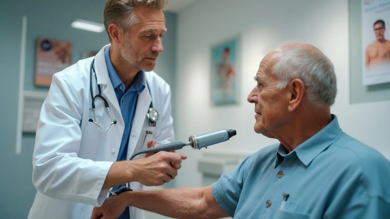
Dermoscopy is a handheld, magnifying device that uses polarized or non‑polarized light to reveal subsurface structures invisible to the naked eye. By enhancing pigment patterns, vascular architecture, and keratinous highlights, dermoscopy boosts diagnostic accuracy from about 65% (clinical exam alone) to over 90% for experienced users.
Key Dermoscopic Features of Actinic Keratosis
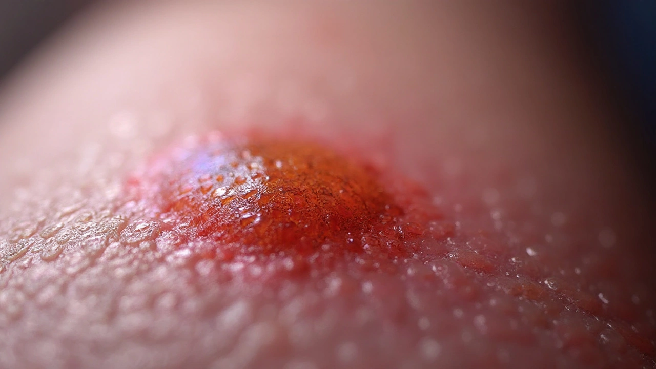
- Red‑strawberry pattern: a background of erythema with dotted or linear vessels.
- White‑yellowish structureless areas indicating hyperkeratosis.
- Targetoid appearance: concentric rings of scaling around a central vascular core.
- Presence of “pseudopods” or peripheral projections, suggesting early invasion.
When these patterns are combined with the lesion’s location and the patient’s UV exposure history, the confidence level for AK rises dramatically.
Comparing Diagnostic Tools: Clinical Exam, Dermoscopy, and Confocal Microscopy
| Feature | Clinical Exam | Dermoscopy | Reflectance Confocal Microscopy (RCM) |
|---|---|---|---|
| Resolution | Macro (≈1mm) | Micro (≈20‑μm) | Cellular (≈1‑μm) |
| Time per lesion | 30‑60s | 1‑2min | 5‑10min |
| Accuracy (experienced clinician) | ~65% | ~90% | ~95% |
| Equipment cost | None | ~$1,200 | ~$30,000 |
| Invasiveness | None | None | None (in‑vivo) |
RCM offers near‑histologic detail but is rarely used in primary care due to cost and training demands. For most dermatology practices, dermoscopy strikes the best balance of accuracy, speed, and affordability.
The UV Connection: Risk Factors You Can’t Ignore
UV radiation is the primary environmental driver of DNA damage in skin cells. Chronic exposure triggers mutations in the p53 tumor‑suppressor gene, laying the groundwork for AK formation. Other risk factors include:
- Fair skin (Fitzpatrick I‑II) \n
- History of sunburns before age 20
- Immunosuppression (organ transplant recipients, chronic steroids)
- Occupational outdoor work
Understanding these factors helps clinicians prioritize high‑risk patients for regular dermoscopic screening.
When Does a Skin Biopsy Become Necessary?
Even the best non‑invasive tools can occasionally miss early invasive changes. A skin biopsy is indicated when dermoscopy reveals atypical vessels, irregular borders, or when the lesion fails to respond to standard therapy after 4‑6weeks. Punch or shave biopsies provide tissue for histopathology, confirming whether the lesion is an AK, SCC in situ, or invasive SCC.
Treatment Options: From Cryotherapy to Topical Agents
Once diagnosed, several effective treatments are available. The choice depends on lesion size, number, location, and patient preference.
Cryotherapy uses liquid nitrogen to freeze the lesion, causing cellular necrosis within seconds. Cure rates range from 85‑95% for isolated AKs. For field‑cancerized skin (multiple lesions), topical agents such as 5‑fluorouracil (5‑FU), imiquimod, or diclofenac gel are preferred.
Photodynamic therapy (PDT) offers excellent cosmetic outcomes for facial AKs but requires specialized equipment and patient cooperation.
Practical Checklist for Clinicians and Patients
- Ask about lifetime sun exposure, occupational history, and prior skin cancers.
- Perform a full‑body skin exam, focusing on sun‑exposed zones.
- Use dermoscopy on any suspicious plaque or papule; look for red‑strawberry pattern, white‑yellow areas, and peripheral vessels.
- Document lesions with high‑resolution photographs for monitoring.
- If dermoscopic features are equivocal, consider a skin biopsy.
- Choose treatment based on lesion count: cryotherapy for isolated spots, topical 5‑FU or imiquimod for field therapy, PDT for cosmetically sensitive areas.
- Educate patients on rigorous sun protection: broad‑spectrum SPF50+, wide‑brim hats, and regular re‑application.
By integrating dermoscopy into routine practice, clinicians can catch Actinic Keratosis before it turns malignant, sparing patients from more invasive interventions.

Frequently Asked Questions
Can Actinic Keratosis disappear on its own?
Rarely. Some small lesions may resolve with diligent sun protection, but most persist or progress. Professional treatment is recommended to eliminate the risk of SCC.
Is dermoscopy painful?
No. The device is handheld and uses light; patients only feel a gentle pressure as the skin is flattened against the lens.
How often should I get screened for AK?
High‑risk individuals (fair skin, outdoor workers, history of skin cancer) should have a full dermoscopic exam annually. Low‑risk patients may be screened every 2‑3years.
What’s the difference between AK and sun‑damaged skin?
Sun‑damaged skin shows diffuse discoloration and dryness, while AK presents as a discrete, rough, scaly plaque that can be felt as a raised area.
Can sunscreen prevent Actinic Keratosis?
Broad‑spectrum sunscreen with SPF30 or higher significantly lowers the incidence of new AK lesions when used daily and reapplied after swimming or sweating.
Is cryotherapy safe for facial AKs?
Yes, when performed by an experienced dermatologist. It may cause temporary hypopigmentation, but cosmetic outcomes are generally good.
When should I consider a skin biopsy?
If dermoscopy shows atypical vascular patterns, if the lesion is ulcerated, or if there is no response to treatment within 4-6weeks, a biopsy is warranted to rule out SCC.
Are there any home‑care methods to lower AK risk?
Regular use of sunscreen, wearing protective clothing, avoiding peak UV hours, and performing self‑exams can all reduce the likelihood of developing new AK lesions.


Michael Waddington
September 25, 2025 AT 14:05