
Pneumocystis Pneumonia Prophylaxis with Immunosuppressants: Who Needs It?
January 14 2026Non‑Invasive Imaging: A Practical Overview
When working with non-invasive imaging, techniques that visualize internal structures without surgical cuts, needles, or radiation exposure. Also known as diagnostic imaging, it lets clinicians spot problems early, plan treatments, and keep patients out of the operating room. Non‑invasive imaging therefore bridges the gap between accurate diagnosis and patient comfort, making it a cornerstone of modern healthcare.
Core Modalities and How They Fit Together
One of the most powerful tools in this family is Magnetic Resonance Imaging (MRI), a method that uses strong magnetic fields and radio waves to create detailed pictures of soft tissues. MRI requires a magnet strong enough to align hydrogen atoms, and the resulting signals are turned into crisp cross‑sectional images. Because it doesn’t use ionizing radiation, MRI is ideal for repeated scans of the brain, spine, and joints. Another staple is ultrasound, high‑frequency sound waves that bounce off organs to produce real‑time images. Ultrasound is portable, inexpensive, and safe for pregnant patients, making it the go‑to for fetal monitoring and bedside assessments. Both MRI and ultrasound fall under the broader umbrella of non‑invasive imaging, showing how the field combines diverse physics principles to meet clinical needs.
Computed Tomography, or CT scan, uses rotating X‑ray beams and computer algorithms to build 3‑D views of bone and dense tissue, adds another layer of capability. While CT does involve low‑dose radiation, its speed and ability to capture fine bone detail make it indispensable for trauma, stroke, and lung evaluations. PET imaging, a functional counterpart, tracks metabolic activity by detecting positron‑emitting tracers; paired with CT or MRI, it creates hybrid images that reveal both structure and function. These modalities illustrate the semantic triple: non‑invasive imaging encompasses structural techniques (MRI, CT) and functional techniques (PET), each requiring specific equipment and expertise.
The rise of hybrid systems, such as PET‑MRI, highlights a key trend: integrating multiple non‑invasive methods to deliver richer diagnostic information. When clinicians combine anatomical detail from MRI with metabolic insights from PET, they can pinpoint tumor boundaries more accurately, guide biopsies, and monitor therapy response without invasive procedures. This synergy demonstrates another triple: advanced imaging technologies enable personalized treatment planning, which in turn improves patient outcomes. As the field evolves, emerging tools like AI‑assisted image analysis and portable handheld ultrasound devices are expanding access, especially in remote or underserved areas.
Below you’ll find a curated selection of articles that dive deeper into these techniques, explore their latest research, and offer practical guidance on choosing the right imaging approach for specific health concerns. Whether you’re a patient curious about what a scan involves or a professional looking for up‑to‑date insights, the posts ahead cover the full spectrum of non‑invasive imaging.
 25 Sep
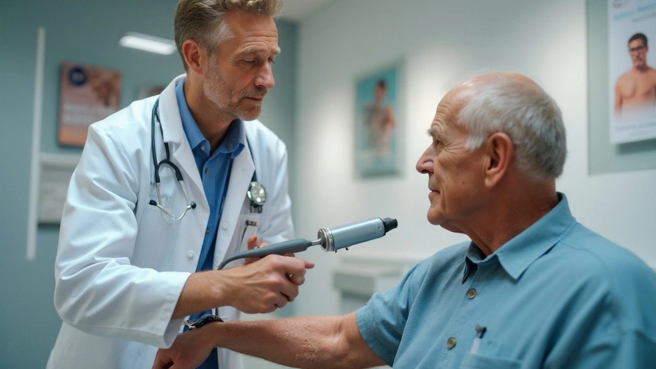
Actinic Keratosis: How Dermoscopy Improves Diagnosis
Learn how dermoscopy helps detect Actinic Keratosis early, its link to skin cancer, risk factors, diagnostic tools, and treatment options in a clear, practical guide.
Read More...